Overview of the technique
STORM is a single molecule localisation microscopy (SMLM) technique. It localises individual molecules and allows achieving resolutions below the Abbe limit. This is achieved by labelling the molecules of interest with a fluorophore (such as GFP) and activate the fluorophores by laser illumination. After being initially fluorescent, they quickly go into a dark state. Over time, they stochastically reappear in small numbers at a time, and their image can be fitted with a Gaussian model. A localisation accuracy of down to 10nm can be achieved. The fitted observations are used to reconstruct a super-resolved image.
The axial position is measured by modifying the point spread function (PSF) to a double helix. The image of the PSF on the camera depends then on the position of the fluorophore relative to the focal plane.
Applications
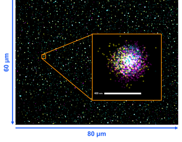
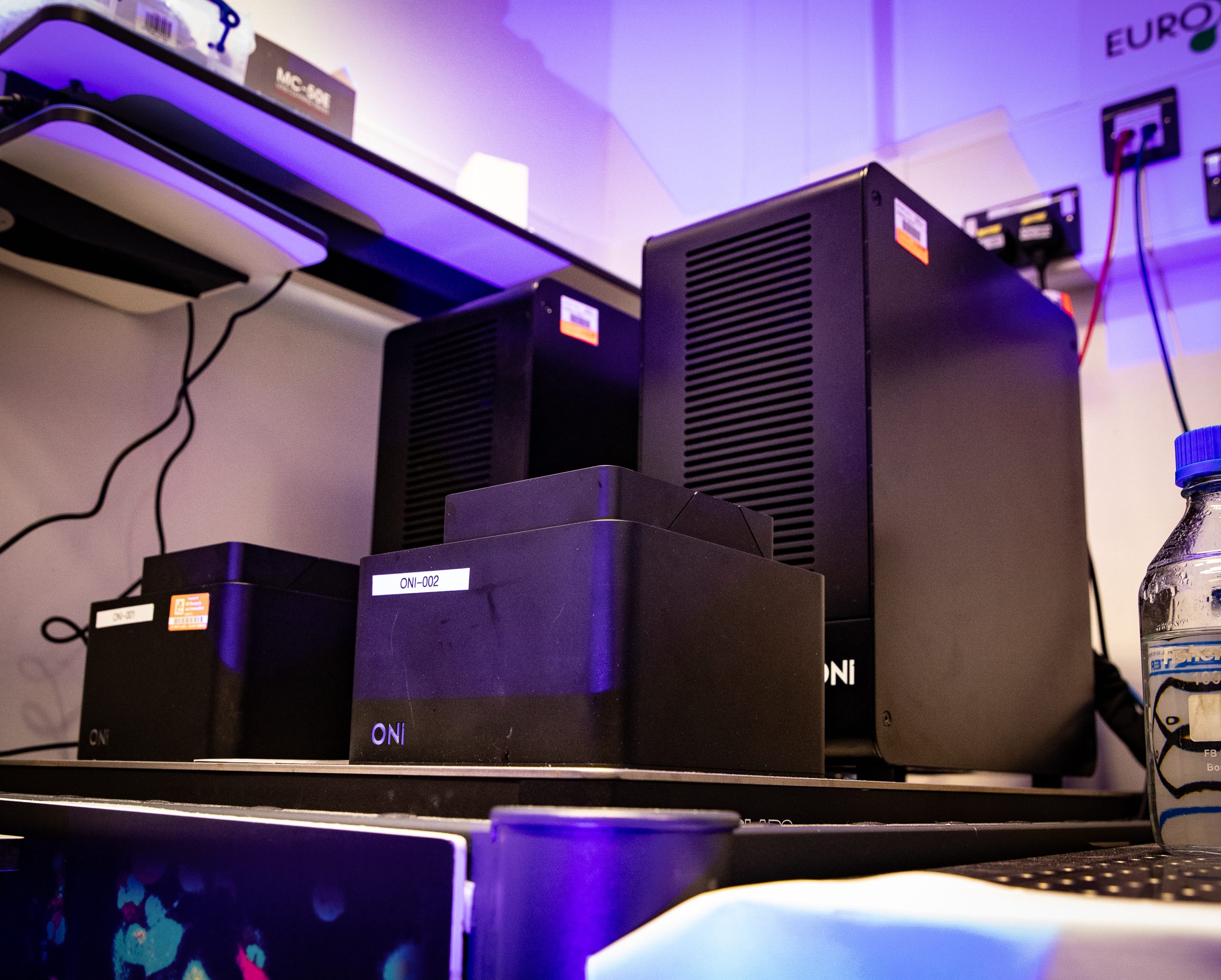
Using dSTORM, researchers can visualize EVs at nanometer resolution, identify key biomarkers, and study their size, morphology, and cargo. The ONI Nanoimager with the EV Profiler 2 kit and AutoEV software streamlines this workflow—combines imaging, and analysis to deliver rapid, high-accuracy insights into EV populations, with Octopus-developed pipelines offering even deeper analysis of heterogeneity.
Exosomes and other extracellular vesicles (EVs) are vital for cell-to-cell communication. They can cross barriers like the blood–brain barrier and enter cells with high specificity, making them strong candidates for drug delivery and diagnostics. Similarly, lipid nanoparticles (LNPs) are lipid-based carriers that protect drugs or genetic material (e.g., mRNA) and deliver them efficiently by merging with cell membranes. Both systems are central to advances in vaccines, gene therapy, and personalised medicine.